External Anatomy of Fishes:
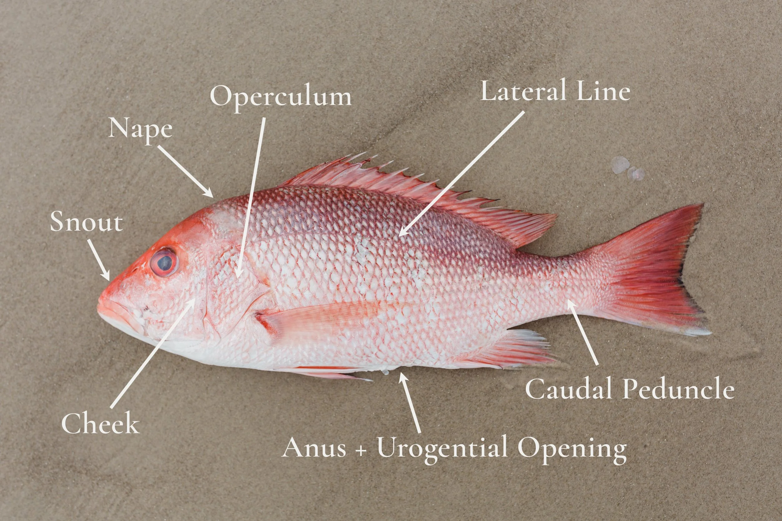
Red Snapper, Lutjanus campechanus
The Red Snapper pictured here was caught locally on Padre Island, TX, using a hook and line, in February, 2024. All images and graphics © Sara Krebsbach 2024.


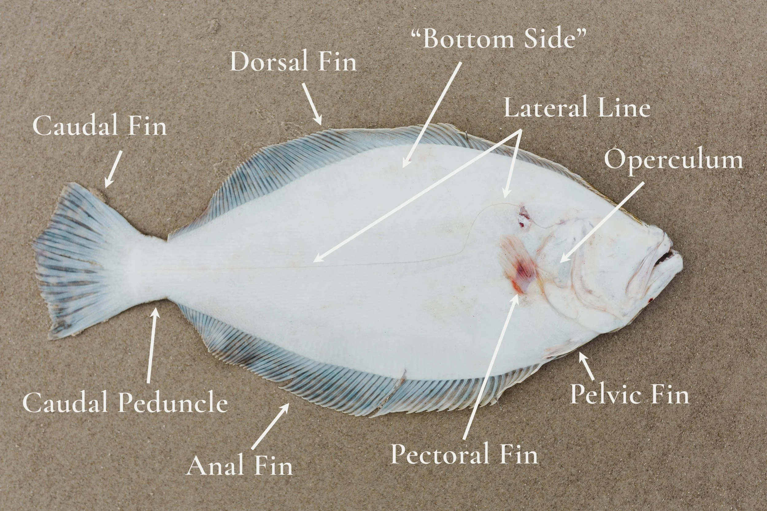
Southern Flounder, Paralichthys lethostigma
The Southern Flounder pictured here was caught locally on Padre Island, TX, using a hand gig, in February, 2024. All images and graphics © Sara Krebsbach 2024.


Lined Seahorse, Hippocampus erectus
The female Lined seahorse pictured here was photographed at Aquarium of Niagara, NY, in April, 2019. All images and graphics © Sara Krebsbach 2024.


American Eel, Anguilla rostrata
The American eel pictured here was shipped to the author in February, 2024, by Captain Brett Stone of No Fly Zone Fishing, in East Dennis, MA. The elver eel was harvested by a licensed eel harvester in coastal Maine, and raised at the aqua-farm, AmericanUnagi, in Waldoboro, ME. All images and graphics © Sara Krebsbach 2024.


Cownose Ray, Rhinoptera bonasus
The male Cownose ray pictured here washed up on Padre Island National Seashore, TX, in February, 2021. All images and graphics © Sara Krebsbach 2024.


Sandbar Shark, Carcharhinus plumbeus
The sharks pictured here were photographed at Texas State Aquarium, TX, in February, 2024. All images and graphics © Sara Krebsbach 2024.


Fish Body Shapes:

All images and graphics © Sara Krebsbach 2024.
Fish Mouth Positions:

All images and graphics © Sara Krebsbach 2024.
Caudal Fin Shapes:

All images and graphics © Sara Krebsbach 2024.
Glossary:
Ampullae of Lorenzini: Electroreceptive organ covering the head and snout area of Chondrichthyes (sharks, skates, rays, and chimaeras), as well as several species of primitive bony fish. Used for detecting electrical fields of other aquatic organisms.
Anal Fin: Fin on median line behind the anus/vent.
Anterior: Directionally toward the head.
Anus: Terminal opening of the alimentary (digestive) canal. Also called a ‘vent’.
Caudal Fin: Tail fin.
Caudal Peduncle: Region connecting the dorsal and anal fin insertion points to the base of the caudal fin.
Cephalic lobes: Used for feeding and communication, and contain electrosensory pores. Derived from anterior pelvic fins. Present in several pelagic species of Batoid rays. Read more.
Cheek: Region of the head ventral and posterior to a fish’s eye.
Claspers: External intromittent copulatory organs of male Chondrichthyes (sharks, skates, rays, and chimaeras), derived from the pelvic girdle. Also called ‘mixopterygia’.
Coronet: ‘Crown-like’ spine seen dorsally on head of Seahorses. Thought to be used to amplify clicking noises used in communication and to attract mates. Coronets are unique to each individual, as a fingerprint is to a human.
Countershading: Camouflage technique seen in many fish species. The dorsal part of the fish is dark in color so that when seen from above, the fish blends in with the bottom environment. The ventral part of the fish is light in color so that when seen from below, the fish blends in with the light entering the water column from above.
Dorsal: The top/upper part of the body.
Dorsal Fin: Fin(s) on the top of the body.
Gill Arch: Bony arches where the gills attach.
Gill Filament: Threadlike projections along the posterior edge of the gill arch, facilitating gas exchange.
Gill Raker: Bony or cartilaginous element on the anterior of the gill arch, opposing the gill filaments.
Gill slits: Openings to gills in species which lack an operculum.
Gills: Respiratory organs for extraction of oxygen from water, and for excretion of carbon dioxide, ammonia, and urea.
Labial Fold: A small fold on the lips at each corner of the mouth.
Lateral Line: Sensory system composed of neuromasts, pores and canals that run throughout the length of a fish’s head and body; used to detect vibrations and water displacement.
Nape: Dorsal area just posterior to the head.
Nares: Nostrils
Nictitating Membrane: A moveable a ‘third eyelid’. Used to protect the eye when attacking prey or other circumstances where protection is warranted.
Operculum: Plate-like structure covering the gills.
Ovipositor: An egg laying organ in female fish; A tube-like egg duct allowing a female to deposit her eggs outside of her body.
Pectoral Fins: Anterior or or most dorsally located of the paired fins.
Pelvic Fins: Paired fins posterior or ventral to the the pectoral fins.
Posterior: Directionally toward the tail.
Precaudal Pit: A small pit on the posterior portion of the caudal peduncle, just anterior of the caudal fin.
Snout: Area of the head between the tip of the upper jaw and the anterior margin of the orbit.
Soft Ray: Bony element that supports the fin; bilaterally paired, segmented, and flexible; used in undulatory movements.
Spine: Bony element that supports the fin; unpaired and unsegmented.
Spiracle: Openings posterior to the eyes in Elasmobranchii (sharks, skates, and rays). Used for respiration in some species. In others, gill lamellae in the spiracle oxygenate blood that is utilized by the eyes and brain.
References:
Castro, J.I. (2011). The Sharks of North America. Oxford University Press.
Freret-Meurer, N. V., Andreata, J. V., & Alves, M. A. S. (2013). Seahorse fingerprints: A new individual identification technique. Environmental Biology of Fishes, 96(12), 1399–1405. https://doi.org/10.1007/s10641-013-0118-6
Hastings, P. A., Walker, H. J., & Galland, G. R. (2014). Fishes: A guide to their diversity. University of California Press.
Helfman, G. S., Collette, B. B., Facey, D. E., & Bowen, B. W. (2023). The diversity of fishes (3rd ed.). Wiley-Blackwell.
Mulvany, S., & Motta, P. J. (2013). The morphology of the cephalic lobes and anterior pectoral fins in six species of batoids. Journal of Morphology, 274(9), 1070–1083. https://doi.org/10.1002/jmor.20163
Ruschenberger, W. S. W., Milne-Edwards, H., & Comté, A. (1846). Elements of Herpetology, and of Ichthyology: Prepared for the Use of Schools and Colleges. Grigg & Elliot.